
IPM Take
Personalised prevention requires identifying risk before established damage becomes apparent. This study suggests that retinal vascular findings could complement eye pressure and other clinical information when assessing future glaucoma risk. However, an association is not yet a validated prediction tool, and the findings do not justify treating cholesterol or uric acid specifically to prevent glaucoma. The next step is to test whether these additional markers improve risk prediction sufficiently to inform practical, equitable screening pathways.
Executive Summary
A retrospective study published online in the American Journal of Ophthalmology followed 17,212 Korean adults without glaucomatous structural changes at baseline. Over a mean of 13 years, 399 participants, or 2.3%, developed reproducible structural changes consistent with glaucoma. Retinal arteriosclerosis was associated with a higher subsequent hazard of this outcome after adjustment for other measured factors: hazard ratio 1.459, with a 95% confidence interval of 1.247–1.708. Older age, higher intraocular pressure, serum uric acid and total cholesterol were also associated with conversion. The findings support further investigation of combined ocular and systemic risk assessment. They do not establish causation, demonstrate visual-field deterioration or validate a screening algorithm for routine clinical use.
Why it matters
- Clinicians: Retinal vascular findings may warrant further investigation as part of a broader glaucoma risk profile. The study does not establish a threshold for referral, a monitoring interval or an indication for treatment.
- Public authorities: The findings provide a research rationale for assessing whether existing retinal photographs can support more targeted preventive pathways. Implementation would require evidence of predictive performance, resource implications and patient benefit.
- Patients and advocates: A possible risk marker is not the same as a glaucoma diagnosis. The study does not show that lowering cholesterol or uric acid prevents glaucoma, and its results should not be used to encourage self-directed treatment.
A retinal photograph may contain more information about future eye health than the finding it was originally taken to assess.
In a large Korean health-screening cohort, researchers found that retinal arteriosclerosis-arterial changes identified in photographs of the back of the eye- was associated with later structural changes consistent with glaucoma. The relationship remained after adjustment for other measured factors.
The finding points towards a broader approach to risk assessment. It also raises a question: when does an observable association become useful enough to change preventive care?
Following risk over time
The study, published online in the American Journal of Ophthalmology on 27 August 2026, analysed 17,212 Korean adults attending the Seoul National University Hospital Healthcare System Gangnam Center. Participants had no evidence of glaucomatous structural change at baseline and had follow-up retinal photographs at least ten years later.
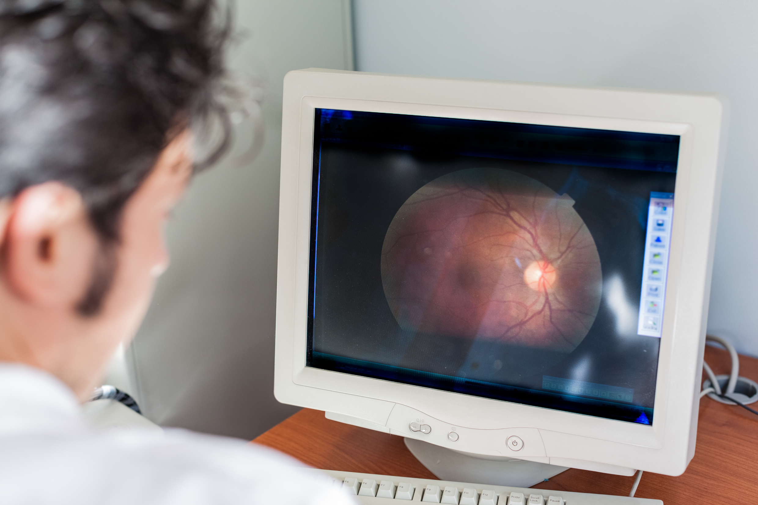
Researchers reviewed serial fundus photographs: images showing the retina and optic nerve region at the back of the eye. Their outcome was the development of definite, reproducible structural changes consistent with glaucoma in eyes whose baseline findings had been normal.
Over a mean follow-up of 13 years, 399 participants developed this outcome, representing 2.3% of the cohort. The incidence rate was 180.2 cases per 100,000 person-years, a measure that accounts for participants’ accumulated follow-up time.
These results describe new structural findings in a selected health-screening population. They should not be read as an estimate of glaucoma incidence in every population.
What the vascular finding means
Retinal arteriosclerosis was independently associated with subsequent structural conversion, with a hazard ratio of 1.459 and a 95% confidence interval of 1.247–1.708. This indicates a higher relative hazard during follow-up, not a 45.9-percentage-point increase in absolute risk.
Other associations included older age and higher intraocular pressure, meaning pressure inside the eye. Each additional millimetre of mercury of pressure was associated with a hazard ratio of 1.158, after adjustment for the other variables in the model.
Higher serum uric acid and total cholesterol were also associated with the outcome. The reported hazard ratios were 1.080 per additional milligram per decilitre of uric acid and 1.096 per additional ten milligrams per decilitre of total cholesterol.
These differently scaled estimates should not be ranked as though they measured interchangeable exposures. Nor does statistical adjustment establish that any of the associated factors caused the structural changes.
An association is not a screening rule
The study’s conclusion is appropriately prospective: microvascular changes, eye pressure and systemic metabolic factors may help refine risk stratification. It does not provide a validated calculator or demonstrate how much retinal arteriosclerosis improves prediction beyond established clinical information.
That distinction matters for implementation. A marker can be associated with disease yet add little practical value to decisions about who should be referred or reviewed sooner.
The outcome also requires careful wording. Researchers assessed structural conversion using photographs; the abstract does not report corresponding rates of visual-field loss, the functional impairment measured by tests of what a person can see. Structural conversion should therefore not be presented as equivalent to symptomatic disease or blindness.
The retrospective design introduces further uncertainty. Because inclusion required photographs at least ten years apart, the findings apply to people who remained available for that long-term assessment, rather than everyone initially entering a screening service.
Personalised medicine opportunity
The personalised medicine opportunity is to combine information rather than elevate one new marker above everything else.
The researchers’ model brings together ocular findings, age and systemic measurements. Their results support investigating whether such combinations can identify people whose future risk differs meaningfully despite apparently normal baseline photographs.
A separate Greek study illustrates why better targeting deserves attention. In the Thessaloniki Eye Study, 46 of 1,042 participants initially free of glaucoma developed open-angle glaucoma during follow-up; 27 of those cases, or 58.7%, had not previously been diagnosed.
That study does not validate the Korean vascular marker. It addresses a different population and question but highlights the practical gap that risk-based detection strategies aim to close. Its authors also called for further evaluation of screening cost-effectiveness.
Looking ahead
Before retinal arteriosclerosis becomes a routine component of glaucoma screening, prospective studies should test whether its assessment is reproducible and whether adding it improves prediction across different populations.
Research should also establish what action follows an elevated estimate. A useful pathway needs more than risk classification: it needs a defensible referral threshold, accessible confirmatory assessment and evidence that the resulting care benefits patients.
For now, the Korean findings support further investigation, not a new screening recommendation. They also offer no basis for prescribing treatment to lower uric acid or cholesterol specifically to prevent glaucoma.
The preventive opportunity lies in learning which existing clinical signals genuinely improve decisions and resisting the temptation to turn every association into an intervention.
