
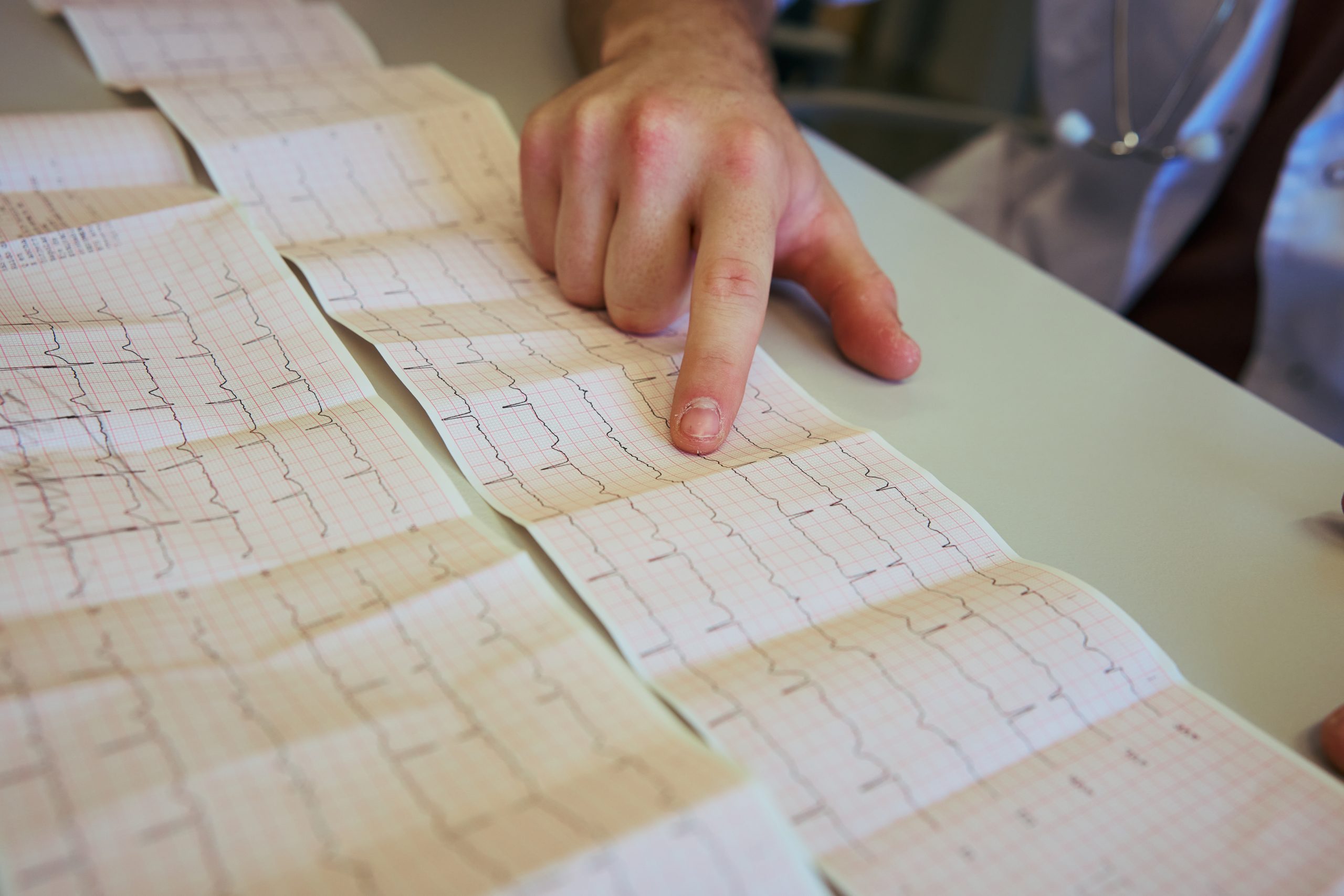
IPM Take
The most important claim about this AI system is not that it can read an ECG in under two seconds. Speed matters little if the result does not change what happens to the patient next.
The more consequential possibility is that a routine test already performed at enormous scale could become a personalised gateway into cardiovascular care. Instead of every patient with possible heart disease entering the same queue for echocardiography, an AI-generated risk signal could identify those most likely to have heart failure or valve disease and move them forward for confirmatory imaging. Researchers at Imperial College London report that the system identified up to 81% of patients with heart failure and up to 90% of those with valve disease in testing involving about 67,000 U.S. patients.
That is a potentially powerful precision-medicine model because it uses routinely collected physiological data to differentiate patients before expensive specialist investigation begins. Yet it also creates an uncomfortable policy question: if AI decides whose scan becomes urgent, the algorithm is no longer simply reading an ECG. It is helping allocate a scarce healthcare resource.
That means accuracy is only the beginning of the evaluation.
Executive Summary
Researchers at Imperial College London’s National Heart and Lung Institute have developed artificial intelligence models capable of extracting patterns from standard electrocardiograms that are not normally visible to clinicians. The technology has been trained on millions of ECGs and is being developed commercially through Imperial spinout Cardiovolt.ai. The initial clinical focus is identifying hidden heart failure and valvular heart disease so that patients at higher probability of disease can be prioritised for echocardiography.
At ESC Congress 2026 in Munich, the team reported testing involving approximately 67,000 U.S. patients, in which the AI identified up to 81% of patients with heart failure and up to 90% of those with valvular heart disease. The analysis can be produced in less than two seconds. The technology is not intended to replace echocardiography or independently confirm either diagnosis.
The clinical concept is therefore one of risk-based triage. A person whose routine ECG contains an AI-detected signal of structural disease could receive faster echocardiography, while patients at lower predicted risk could remain within the standard pathway. A related Imperial programme is already exploring how AI-enhanced ECGs could be integrated into clinical practice, and the technology is moving toward real-world NHS evaluation.
For personalised medicine, the attraction is clear, but implementation will require prospective evidence that AI-guided triage reduces diagnostic delay and improves outcomes without generating excessive false positives or widening existing inequalities in access to cardiac imaging.
Why it matters
- HTA bodies: Assessment should move beyond algorithmic performance and ask whether AI-guided ECG triage reduces time to diagnosis, improves treatment initiation and generates enough clinical benefit to justify additional downstream imaging. Sensitivity, specificity, positive predictive value and performance across different populations will matter more than processing speed.
- Payers: Re-using an ECG that is already being performed could be inexpensive compared with creating an entirely new screening programme, but widespread AI deployment may increase demand for echocardiography by revealing previously unsuspected disease. The cost question is therefore not only what the algorithm costs, but what happens after it flags a patient.
- Industry / innovation partners: The model illustrates a broader opportunity in precision diagnostics, where AI extracts new biomarkers from existing clinical data rather than requiring a new test. Commercial success will depend on prospective validation, interoperability with hospital ECG systems and evidence that the risk prediction leads to better patient pathways rather than simply more alerts.
The electrocardiogram has been part of cardiovascular medicine for more than a century, yet its conventional clinical role remains relatively narrow. It records the electrical activity of the heart and is particularly useful for recognising rhythm abnormalities, conduction disturbances and signs associated with myocardial infarction, while structural conditions such as heart failure and valve disease generally require additional investigation, most importantly echocardiography.
Researchers at Imperial College London are testing whether artificial intelligence can extract substantially more information from those same electrical signals.
Their system has been trained on millions of ECG recordings and is designed to recognise patterns that are too complex or subtle for conventional visual interpretation. According to results reported at ESC Congress 2026, analysis takes less than two seconds and, in testing involving approximately 67,000 patients in the United States, identified up to 81% of patients with heart failure and up to 90% of those with valvular heart disease.
The language around the research has understandably focused on a “superhuman” ECG, but the more useful policy interpretation is considerably less futuristic. The system could become a triage layer between a routine test and specialist imaging, using an individual’s ECG to estimate who is most likely to need an echocardiogram quickly.
Personalisation begins before treatment
Personalised medicine is often discussed in terms of choosing a drug according to a genomic mutation, molecular biomarker or disease subtype, but personalisation can begin much earlier in the pathway. It can determine who receives further investigation, how urgently that investigation occurs and which diagnostic pathway a patient enters.
That is particularly relevant to heart failure and valve disease because delayed diagnosis can postpone treatments that are more effective when introduced before advanced deterioration. Yet echocardiography requires trained staff and specialist capacity, and demand can exceed supply.
The Imperial model proposes a different use of the ECG. Rather than treating everyone who has undergone the test as diagnostically equivalent, the AI would calculate information from the patient’s individual electrical pattern and flag those with a higher probability of structural disease. Imperial researchers have described the potential to run the model across ECGs already being performed in hospitals, including those ordered for unrelated reasons, creating an opportunity for incidental early detection.
For health systems, this is attractive because the underlying diagnostic infrastructure already exists. An ECG takes seconds to record, is inexpensive and is among the most commonly performed medical tests worldwide. Adding an algorithm could therefore extend the value of an established test without asking every patient to undergo a new screening procedure.
That is the personalised-medicine promise: extracting more clinically relevant information from data the health system already has.
A better algorithm can still create a worse pathway
The difficult questions begin once the AI produces its result.
The researchers are clear that the system cannot independently diagnose or exclude heart failure or valve disease. A positive signal still requires clinical assessment and, in most cases, confirmatory echocardiography.
That means implementation could produce two very different outcomes.
In the successful version, high-risk patients are identified earlier, echocardiography is prioritised according to clinical probability, treatment starts sooner and unnecessary diagnostic delay is reduced.
In the unsuccessful version, the algorithm produces thousands of additional flags, imaging demand rises faster than capacity, low-risk patients are pushed into further testing and waiting lists simply move from one part of the system to another.
Previous research on AI-based ECG screening illustrates why this distinction matters. A meta-analysis of AI models for valvular disease found a pooled sensitivity of 83% and a very high negative predictive value, but the pooled positive predictive value was only 13%, meaning many positive signals would not ultimately represent valve disease in the populations studied. The review concluded that AI-ECG screening needs to operate alongside clinical judgement rather than as a stand-alone diagnostic system.
The exact predictive values of the Imperial system in different clinical populations will therefore be critical. A tool used in a specialist cardiology clinic, where disease prevalence is high, may perform very differently when applied opportunistically to every ECG performed across an entire hospital.
Population matters too. Models trained on millions of historical ECGs can inherit differences in age, ethnicity, sex, comorbidity and healthcare access found in the data used to develop them. Before AI-generated scores begin determining who moves forward in a diagnostic queue, health systems will need evidence that performance remains reliable across the populations they serve.
That makes this as much a governance problem as a technical one.
ESC itself made artificial intelligence the spotlight of its 2026 Congress, with sessions covering AI-based ECG interpretation, imaging and risk prediction, while emphasising the transition from technology development to safe clinical implementation.
The distinction is important because healthcare has no shortage of impressive algorithms. What it needs are technologies that improve pathways.
If AI can turn the ECG into a personalised early-warning system, it could help move cardiology away from a first-come, first-scanned model toward investigation based more closely on individual disease probability. That could be particularly valuable where echocardiography capacity is constrained.
But precision medicine is not achieved when an algorithm produces a personalised number. It is achieved when that number leads to a better decision for the person behind it.
For AI-ECG technology, the decisive endpoint will therefore not be whether heart disease can be detected in two seconds.
It will be whether the health system can act intelligently in the seconds, days and weeks that follow.
