
IPM Take
The value of identifying a rare disease often begins with avoiding the wrong pathway. This series describes a PLAAT4-associated disorder whose spinal imaging pattern and family history may distinguish it from inflammatory demyelination. The finding is a diagnostic lead, not a treatment breakthrough.
Executive Summary
Investigators studied 26 people from 11 unrelated families at four Chinese tertiary centres. Two recurrent heterozygous PLAAT4 variants were associated with an autosomal-dominant condition marked mainly by progressive leg spasticity. MRI showed variable brain white-matter changes and a distinctive symmetric, long-segment pattern in the spinal cord. A mouse model supported a gain-of-function mechanism. Plasma lipid changes were proposed as possible progression markers but are not validated for clinical monitoring.
Why it matters
- Patients and families: A specific diagnosis can clarify the care pathway and inform genetic counselling.
- Clinicians: The combination of progressive spasticity and distinctive spinal imaging may warrant specialist genetics review.
- Diagnostics / pathology: Variant interpretation must be matched to imaging, symptoms and inheritance rather than a scan alone.
- Payers: Access to appropriate genetic assessment may prevent prolonged diagnostic work-ups and potentially unnecessary treatment.
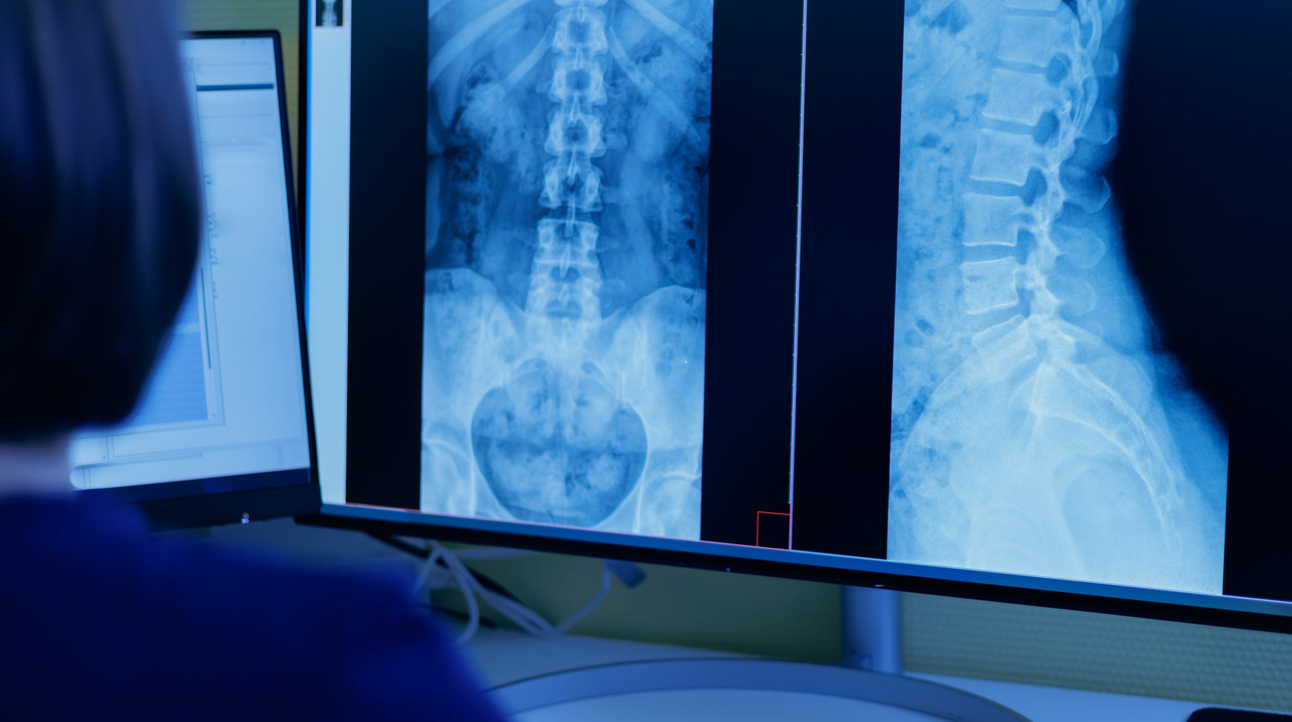
The PLAAT4 case series in JAMA Neurology describes patients whose progressive leg stiffness and white-matter abnormalities could be mistaken for a more common inflammatory disorder. The recurring clue was a symmetric, long-segment, “girdle-like” pattern of spinal cord changes. In some presymptomatic variant carriers, spinal changes appeared before brain involvement.
Researchers combined clinical assessment and whole-exome sequencing with functional experiments. Recurrent PLAAT4 variants appeared across unrelated families, while an engineered mouse model supported the proposed demyelinating mechanism. These complementary findings make the disorder more recognizable, although the clinical series is small and comes from specialist referral centres.
Recognition could help a patient reach the right diagnostic service and avoid treatment based on an incorrect inflammatory diagnosis. It still requires careful interpretation: neither a suggestive MRI nor a genetic variant in isolation is enough to establish the condition. The reported plasma lipid associations are an avenue for further research, not a ready-made progression test.
